First-trimester screening is performed from the beginning of the 11th week of pregnancy (11W + 0D) to the end of the 13th week of pregnancy (13W + 6D) when the fetal length (CRL) is in the range of 45 to 84 mm. In the first trimester of screening, the risk of three common chromosomal abnormalities including Down's syndrome (trisomy 21), Edward's syndrome (trisomy 18),andd Pato's syndrome (trisomy 13) in the fetus is calculated. In all three mentioned syndromes, mental retardation is one of the symptoms. The results of the first-trimester screening show us the probability of a pregnancy with Down syndrome, trisomy 18, or trisomy 13, which is called the risk ratio in the screening report. The accuracy of this screening is almost 94% and false positive cases are 8.4%.
The following items are used in the first-trimester screening:
-
Background risk
-
Ultrasound indicators
-
Biochemical or laboratory indicators
Background risk:
In the screening consultation, the information of this section is completed by drawing a family tree and taking family and personal history. In this section, the age of the mother is one of the important factors. In the case of pregnancy with a donated egg, the age of the egg donor also becomes important. Another important thing is the child's previous history of Down syndrome. Other relevant factors in the screening consultation include ethnicity, type of pregnancy in terms of number of fetuses (if known), method of pregnancy (natural or using assisted reproductive methods), number of pregnancies, number of children, history of diabetes, and smoking, as well as cases Another question is also asked. In this consultation, the goals of screening and the difference between the goals of screening tests and diagnostic tests are explained so that mothers have a correct understanding of the concept of positive screening, which does not necessarily indicate fetal infection.
- Ultrasound indicators:
- The thickness of the back of the neck of the fetus (NT: Nuchal Translucent): the increase in the thickness of the soft tissue behind the neck is the most well-known and widely used marker that can be detected very early and is of great importance in screening.
- Fetal Heart Rate (FHR)
- Checking the presence of nasal bone (NB: Nasal Bone): The lack of formation or hypoplasia of the fetal nasal bone in pregnancy is one of the helpful symptoms
- Investigating right atrioventricular valve failure of the fetal heart (TR: Tricuspid Regurgitation): its observation strengthens the possibility of the presence of these chromosomal disorders in the fetus.
- Examining fetal abdominal vein blood flow (DV PI: Ductus Venosus Blood Flow): Abnormality of DV also increases the possibility of these diseases in the fetus.
- Biochemical or laboratory indicators:
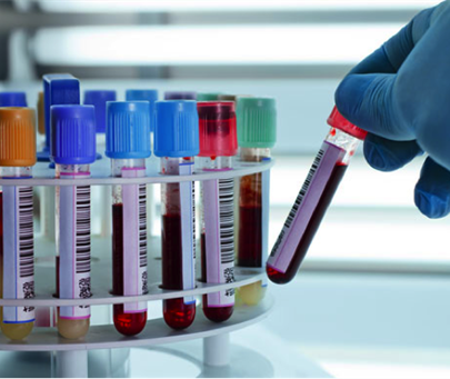
Pregnancy Associated Plasma Protein A (PAPP-A): The origin of this substance is the placenta, and its concentration increases steadily during a normal pregnancy. It has been proven that a significant reduction of this substance is related to chromosomal disorders in the fetus, especially Down syndrome.
Free β hCG: This hormone is secreted during pregnancy first by the origin of the corpus luteum and then by the chorion and placenta. In a pregnancy with a fetus with Down syndrome, its value is higher than expected, and it decreases in trisomy 13 and 18.
The screening results of the first trimester are presented in one of the following three ways:
- High-risk group (Screen Positive): It is recommended to consult and perform amniocentesis or CVS diagnostic tests. (High risk in screening does not mean definite infection)
- Low-risk group: continuation of usual pregnancy care is recommended. (Low risk does not mean any infection, it means a low probability of infection)
- Intermediate Risk Group: In HopeGeneration Medical Institute, according to the FMF protocol, in cases where the risk is intermediate and the mother's age is less than 40 years and the NT size is less than 3 mm, the measurement of other ultrasound markers including the nasal bone (NB: Nasal Bone) and tricuspid regurgitation (TR: Tricuspid Regurgitation) and ductus venosus (DV PI: Ductus venosus PI) abnormalities are checked and finally the fetus is placed in the low-risk and high-risk groups. In the rest of the people who report an Intermediate screening result, it is recommended to perform Cell-Free Fetal DNA (NIPT).
In twins, screening criteria are the same as in singleton pregnancy, but in more than twin pregnancies, biochemical indicators are not used in screening.

